Scientists shrank mice's brains and replaced the missing tissue with human 'organoids'
In a new study, scientists reared lab mice that were missing sections of their brains and then replaced the missing tissue with human cells. The experiment represents a step forward in the study of …
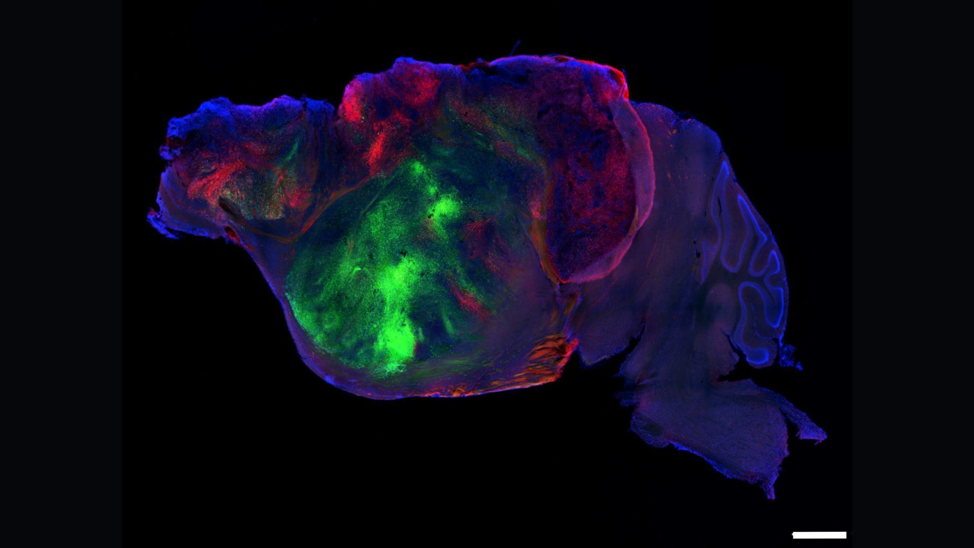
In a new study, scientists reared lab mice that were missing sections of their brains and then replaced the missing tissue with human cells.
The experiment represents a step forward in the study of human brain organoids — tiny models of the human brain grown from stem cells. In the long run, scientists aim to use organoids to better understand how the brain develops and how its structure and function change in the context of disease.
"It is definitely an advance in the field," said Dr. H. Isaac Chen , an associate professor of neurosurgery at the University of Pennsylvania Perelman School of Medicine. Chen was not involved in the current study but has conducted experiments transplanting human brain organoids into rodents' heads .
Organoids offer a window into early brain development that's impossible to observe closely in humans, namely because it unfolds inside developing fetuses. Although organoids are not perfect re-creations of full-size human brains , scientists think organoids are useful models for studying the developing brain in both health and disease.
The model in the new study "certainly creates some pretty interesting options in terms of modeling human neurodevelopment and various types of neurodevelopmental disorders," Chen said. "If you're looking for a model that really allows you to look at larger areas of human neural tissue from a cellular, molecular perspective, I think there's a lot that this model has to offer."
A top-down view of a mouse brain grafted with human brain organoids (labeled in green and red). The scale bar is 0.08 inches (2 mm).
The white lines outline human brain organoids within the mouse's brain tissue. The colors indicate the main direction each nerve fiber is going within the organ: green (top to bottom); red (front to back); and blue (side to side).
An MRI showing human brain organoids (outlined in dashed white lines) and the nerve fibers extending from them. The colors indicate the main direction each fiber is going: green (top to bottom); red (front to back); and blue (side to side).
Read Full Story at Live Science →